Our Space
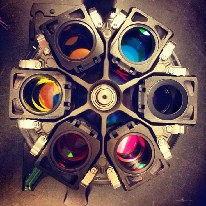
We are in the 7th floor of the Brown building of the Department of Biology at NYU, just one block away Washington Square Park. Our lab has an open plan configuration to foster interactions and collaborations between lab members as well as with members from other research groups. Our research combines experimental and computational tools. On the experimental side, we use microscopy and image analysis, cell culture, microfabrication, metabolite quantification, molecular biology and in vivo studies. As part of the Center for Genomics and Systems Biology we have access to state-of-the-art computational facilities.
Our Hardware
In addition to our cell culture and molecular biology facilities, our lab is implementing and installing several advanced equipment including:
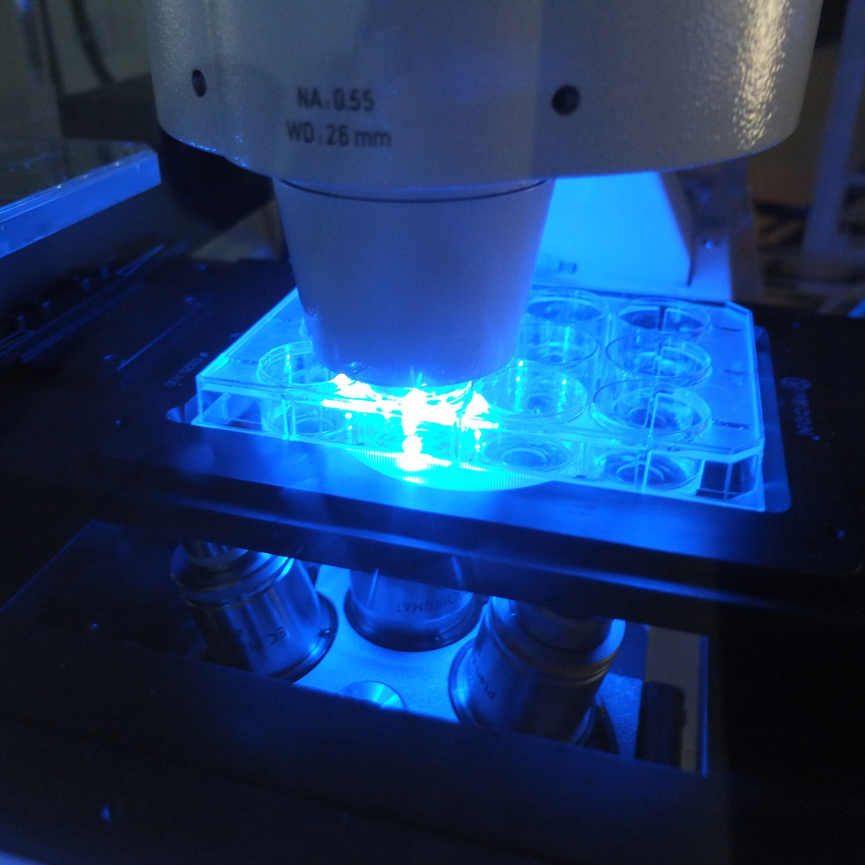
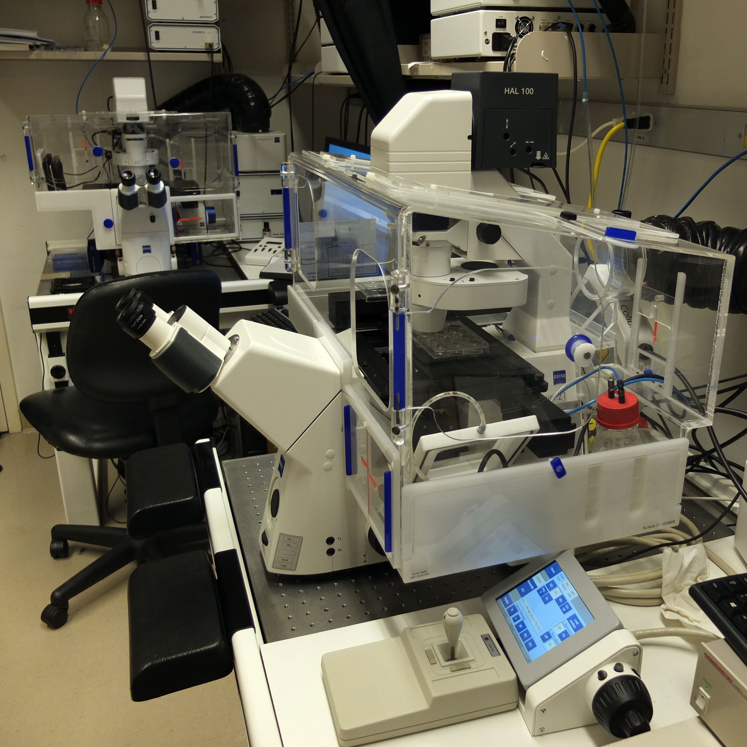
- - Confocal microscopy for live imaging.
- - High content live imaging system.
- - Liquid handling robot.
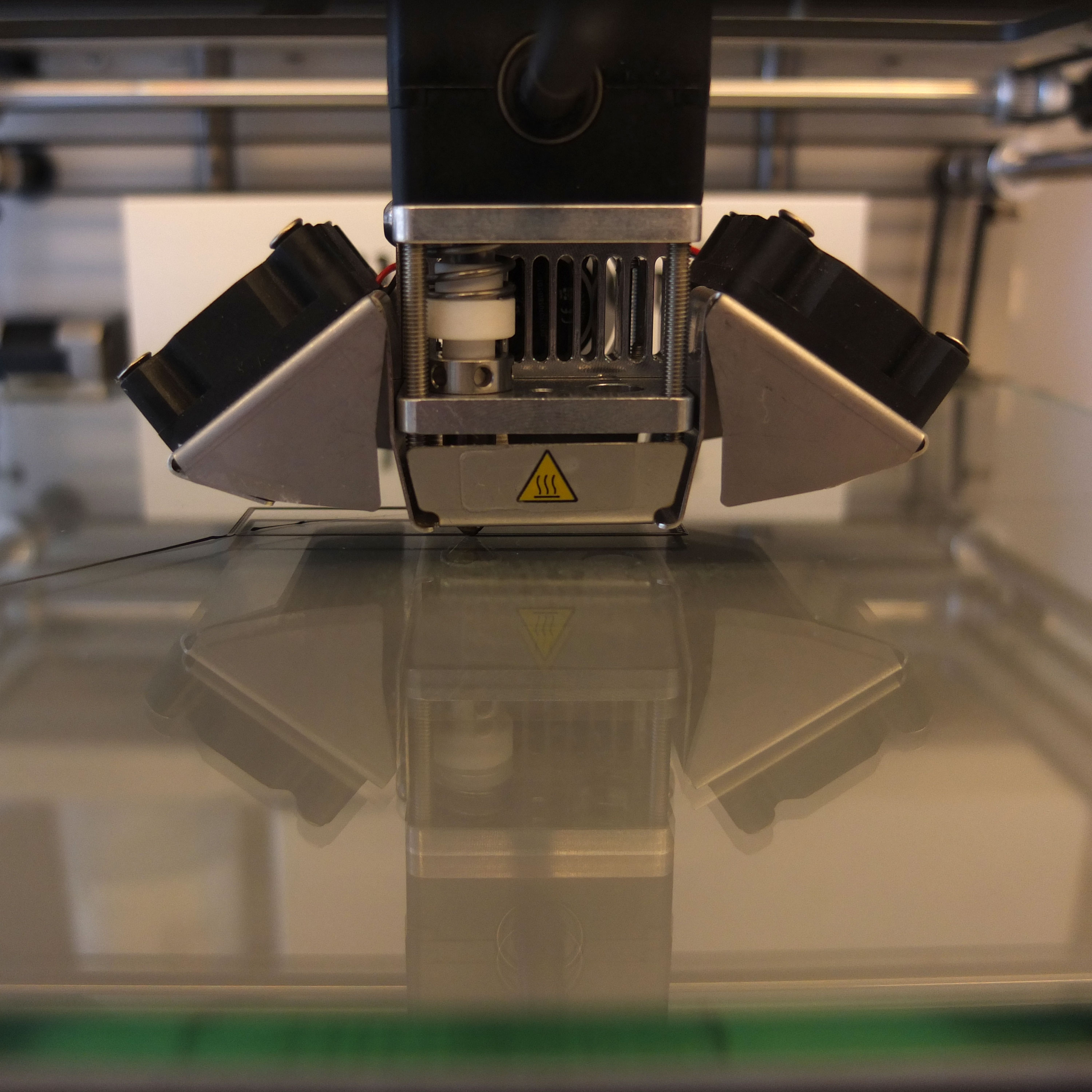
- - 3D printer and laser cutting technologies.
- - Access to metabolomic facilities.
- - Access to animal facilities.
- - Computer workstations and access to high-performance computing facilities.
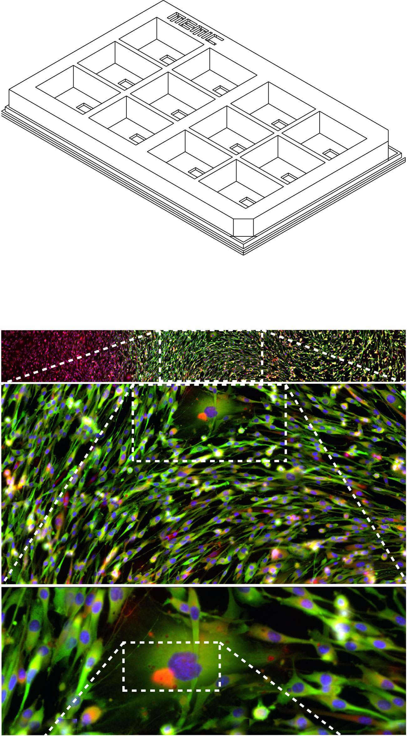
The MEMIC
Cells within tumors do not experience the same environments. Levels of extracellular metabolites vary according to proximity to the vasculature, where oxygen and nutrient levels are normal in perivascular regions but conditions turn gradually more ischemic in distal regions. This is not exclusive to tumors and we are exploring other systems where this may be occurring.
We have shown that these graded environments have functional consequences and modulate tumor progression. But how to study this problem? In an in vivo setting, is hard to untangle the effect of metabolic gradients from other variables, such as cell signaling and recruitment of cells. On the other hand, traditional in vitro culture systems allow controlling for some of these variables but they are spatially homogeneous without major differences in metabolite distribution. To overcome these limitations, we developed a microphysiological system that mimics the microenvironment of tumors using laser-cutting and 3D-printing technologies. This system, called MEMIC (MEtabolic MIcroenvironment Chamber), attempts to emulate metabolic gradients formed between cells in close and distant proximity to the blood supply. Briefly, cells are cultured in a small volume (~70uL) connected through a small slit to a larger chamber (2mL) filled with fresh media. The media in the small chamber, which is conditioned by metabolites consumed and released by the cells, is exchanged by passive diffusion with fresh media through the slit. This exchange generates spontaneous extracellular gradients where the medium composition at the slit is equal to fresh media but becomes progressively ischemic with distance.
Read more about our work using the MEMIC system in our publications page. Download here the .stl files required for printing MEMICs using a 3D printer. If you use our MEMIC please cite our paper where we describe it.
Click here to see the current state of our 3D printers (NYU intranet required).

